
3D Medical Imaging Software for Medical Devices – Compliance & Workflow Guide
Any sufficiently advanced technology is indistinguishable from magic.” Arthur C. Clarke’s Third Law, Profiles of the Future, 1973.
Are you aware that in today’s world you can visualize the opening and closing of a patient’s mitral valve from a 3D perspective? Do you think this is magic? In fact, it’s not magic but an explosion of technology in 3D Imaging Software that enables healthcare professionals to see not only the complex structures of the human body but also their real-time function, enhancing diagnosis, treatment planning, and patient outcomes.
After all, what brings a patient to the doctor? Malfunction. Malfunction causes pain and organ failure. It can even cause death. Imagine seeing actual anatomy, physiology, and malfunction in action! Imagine the diagnostic and therapeutic possibilities.
Elexes has supported 3D medical imaging software regulatory clearance for clients in Europe & US. As an RA Director in a medical software company, understanding 3D imaging software regulation is crucial – this guide helps you navigate the path to clearance.
3D software is quickly becoming the standard of care for the following:
⦿ Presurgical evaluation and planning: incorporating the aesthetics into the repair in dental and cosmetic specialties
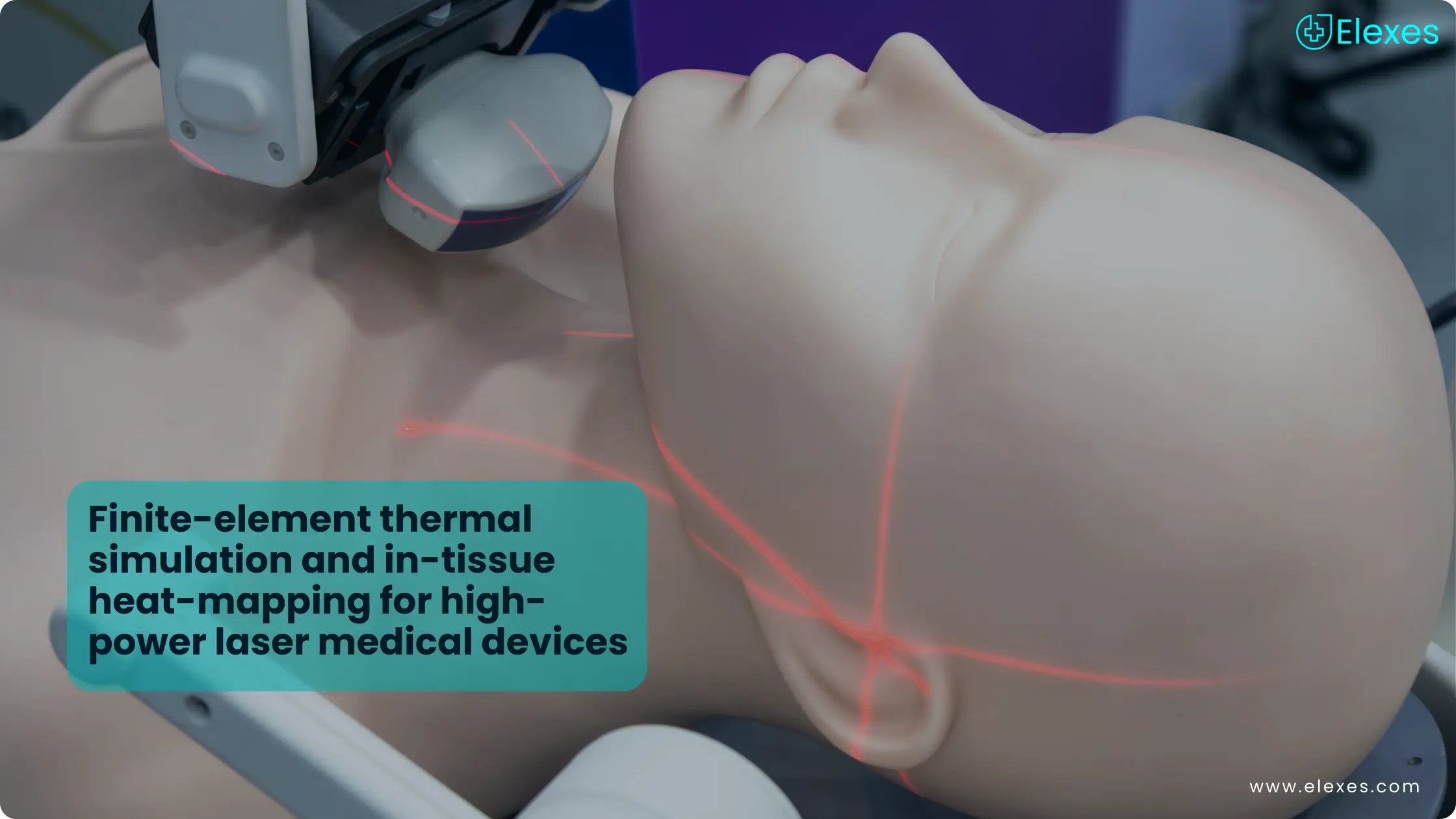
⦿ Surgical/treatment guidance: hand-in-hand with today’s evolving robotic surgery to further reduce recuperation and hospitalization
⦿ Assessment of treatment response: seeing before and after in 3D gives better accuracy for both
⦿ Fusion technologies: the human body does not like extra holes, so visualization for more accurate localization of the problem area and accurate biopsy targeting in 3D, assures there will be fewer of them
⦿ Effective communication of information: data compression that can make imaging travel between doctors and facilities timely and efficiently
According to Clarke’s Third Law, this really is magic! Have a look at these groundbreaking software magicians:

⬩ Surgical Planning Preview® Treatment Planning Software by M2S
The Preview software for 3D modelling can help visualize highly angulated, fenestrated, branched, tortuous vasculature and contrast and non-contrast data. The software demonstrates interactive 3D models for endovascular and aneurysm repair.
⬩ 3D Surgery™ by Dolphin
Orthodontic and implant surgery can now integrate aesthetics with the function like never before.
Orthopedic pre-operative planning and templating software for joint surgical therapy, replacement and pediatric deformity strategies.
⬩ Treatment Planning
Eclipse Treatment Planning System by Varian Medical Systems
Being an integrated and comprehensive treatment planning system, Eclipse renders precise targeting in radiation treatment techniques such as photon, FFF beam, proton and electron external beam, brachytherapy and Cobalt. This system includes simplified data settings and easy drag and drop functionality that can increase the productivity for clinicians. It helps clinicians to create, import and optimize plans prior to the treatment.
⬩ Diagnosis
Allura 3D-RA by Philips
Allura 3D-RA provides high-speed, high resolution 3D visualization of vascular pathologies anywhere in the body. When used with Philips Allura FD systems, it can even cover anatomy including cerebral, abdominal and peripheral vasculature. It features the Reconstructive Zoom Technique that enables high-resolution 3D images to be created for smaller fields of view.
⬩ Visualization and Sharing X-FRAME CCD by Medical Expo
This digital radio/fluoro system with its high resolution, sensitivity and power processing tools is ideal for visualization, acquisition and sharing of sophisticated dynamic studies.
The technology that these are based on are the DICOM and JPEG 2000 which are state-of-the-art image compression standards for storage and transmission of medical images.
Delay means worsening patient jeopardy, and JPIP (JPEG 2000 Interactive Protocol) is a compression streamlining protocol that works with JPEG 2000 to produce an image using the least bandwidth required, so as to get imaging to remote locations as quickly as possible.
Validation
Software validation is used to demonstrate that the software will function safely and effectively as intended. This is very critical from a regulatory, clinical and quality systems standpoint. Standards and guidance documents applicable to the 3D imaging software are:
- NEMA PS 3.1 – 3.20 (2011) Digital Imaging and Communications in Medicine (DICOM) Set
- NEMA PS 3.1 – 3.18 (2008) Digital Imaging and Communications in Medicine (DICOM) Set
- IEC/ISO 10918-1 First edition 1994-02-15 Information Technology – Digital Compression and Coding of Continuous-Tone Still Images: Requirements and Guidelines [Including: Technical Corrigendum 1 (2005)]
- IEC 62304:2006 Medical device software – Software life cycle processes
- Guidance for the Submission of Premarket Notifications for Medical Image Management Devices, July 27, 2000
- General Principles of Software Validation; Final Guidance for Industry and FDA Staff, January 11, 2002
- Guidance for the Content of Premarket Submissions for Software Contained in Medical Devices, May 11, 2005
3D Imaging Software Compliance Checklist
FAQs
What class of medical device is 3D imaging software?
3D imaging software is typically classified as a Class II medical device in the US and a Class IIa/IIb under the EU MDR, depending on its intended use and impact on clinical decision-making.
Does 3D imaging software require FDA 510(k) or CE-mark?
Yes, most 3D imaging medical software requires a FDA 510(k) clearance and a CE-mark before clinical use.
What standards apply to 3D imaging medical device software?
Applicable standards include IEC 62304 for the software lifecycle, ISO 13485 for quality management, ISO 14971 for risk management, and IEC 62366-1 for usability engineering.
How do you validate segmentation and STL accuracy for 3D models?
Validation is done by comparing output against ground truth datasets and quantifying geometric deviations using appropriate metrics to ensure segmentation and STL accuracy.
What are best practices for implementing 3D imaging software into a hospital workflow?
Best practices include clinician training, integration with PACS/EMR, and defined QA procedures, validation in the clinical environment, and periodic performance monitoring to ensure consistent results.